What is Histotripsy (HistoSonics)?
Histotripsy, developed by HistoSonics, is an innovative, non-invasive, robotic ultrasound therapy used to destroy cancerous tissue in the liver.
Histotripsy works by delivering high-intensity, focused ultrasound pulses to a targeted tissue area, creating controlled acoustic cavitation. This cavitation forms microscopic bubbles in the tissue, which rapidly expand and collapse, generating strong mechanical forces. These forces physically break apart cellular structures, effectively liquefying the targeted tissue without using heat or making any incisions. The body then naturally absorbs and removes the destroyed tissue.
Indications for Histotripsy (HistoSonics)
Histotripsy is typically indicated to treat tumorous tissue in the liver in:
- Individuals with primary liver cancer
- Individuals with other cancers that have spread to the liver
- Individuals who have not had chemotherapy, immunotherapy, radiation, or surgery
- Individuals whose disease has spread despite previous treatment
- Individuals who are not candidates for other treatment options
What Does Procedure for Histotripsy (HistoSonics) Involve?
Histotripsy procedure for liver tumors is typically performed under general anesthesia and fasting may be required prior to the procedure. In general, the procedure involves the following steps:
- The patient lies on a procedure table similar to that used in imaging studies.
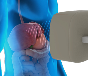
- The HistoSonics Edison™ system (robotic arm + ultrasound probe) is positioned over the body.
- Real-time ultrasound imaging helps localize the tumor in 3D.
- The treatment area is marked and confirmed using software planning tools.
- High-intensity, focused ultrasound pulses are delivered to the target through the skin.
- The focused energy creates microbubble cavitation in the tumor, leading to mechanical tissue destruction.
- Tissue is fractionated into acellular debris while sparing surrounding healthy tissue like vessels and ducts.
- The system automatically adjusts the focal point and depth for uniform ablation.
- The procedure may typically take 30–90 minutes, depending on tumor size and complexity.
What Happens After the Histotripsy (HistoSonics) Procedure?
After a histotripsy procedure, the destroyed tissue is gradually broken down and absorbed by the body through natural healing processes. Since the treatment is non-invasive and non-thermal, most patients experience minimal pain, no scarring, and a quick recovery. There is typically no need for hospitalization, and patients can often return to normal activities within a day or two. Follow-up imaging may be performed to monitor the treatment area and assess the reduction in tumor size or tissue debris. Overall, post-procedure recovery is generally smooth with a low risk of complications.
Risks and Side Effects
Although histotripsy is designed to be a minimally invasive and non-thermal treatment with a favorable safety profile, like any medical procedure, it does carry some risks and side effects. These include:
- Fatigue
- Mild discomfort or soreness
- Low-grade fever
- Bruising or skin irritation
Benefits
Benefits of the histotripsy procedure include:
- No scalpel, incision, or thermal injury
- Fast recovery, often same-day discharge
- Preservation of nearby critical structures
- Real-time visualization during treatment
If you wish to be advised on the most appropriate treatment, please call to schedule an appointment or click to request an appointment online.